Nasal Septum Diagram Labeled : Oral/Nasal Cavity Model - PurposeGames - Posteriorly it meets the concave anterior margins.. It is depressed by the depressor septi nasi muscle. Each image highlights and labels the sinuses in coronal and sagittal view. Septum nasi) separates the left and right nasal cavities. Note that the septum has a hard part (here partly bony and partly cartilaginous) and a soft or mobile part. The nasal septum divides the left and right nasal cavities (fig.
Nasal septal deviations play a critical role in nasal obstruction symptoms, aesthetic appearance of flow diagram for the literature search and overall study selection. Keystone areas preserve along bony cartilaginous junction preserve along nasal floor diagram showing area of l shaped strut. Septum nasi) separates the left and right airways of the nasal cavity, dividing the two nostrils. Download scientific diagram | draf classification for frontal sinus drainage. It extends from the nares anteriorly to the choanae posteriorly and is anteriorly the septal cartilage (or quadrangular cartilage) which approximates a quadrilateral shape.
Nasopharynx; Rhinopharynx from upload.wikimedia.org The nasal septum contains bone and hyaline cartilage. Septal splints of universal size and shape were prepared from silastic sheeting. A deviated septum occurs when your nasal septum is significantly displaced to one side, making one nasal air passage smaller than the other. The label aspergillosis is used for several diverse clinical conditions. The nasal septum is composed of cartilage in the front and bone in the back of the nose. What is nasal cavity definition, what is the function of nasal cavity, role of mucus in nasal cavity, anatomy, structure, nasal cavity bones, labeled diagram. The cartilage also gives shape and support to the outer part of the nose. Nasal septum area was determined in 21 caucasian body donors, and nasal septum thickness was measured in 20 ct scans.
The nasal septum contains bone and hyaline cartilage.
Nasal septum deviation is a physical disorder of the nose, involving a displacement of the nasal septum. Nasal septum is comprised of: The medial wall or septum is frequently more or less deflected from the median plane, thus lessening the size of one nasal cavity and increasing that of the. Posteriorly it meets the concave anterior margins. Advertisements present are clearly labelled and in no way support the. A small part of the nasal roof and. What is nasal cavity definition, what is the function of nasal cavity, role of mucus in nasal cavity, anatomy, structure, nasal cavity bones, labeled diagram. Also note that the floor of the nasal cavity is. The nasal septum contains bone and hyaline cartilage. Each image highlights and labels the sinuses in coronal and sagittal view. The nasal septum is composed of five structures: Smartdraw includes 1000s of professional healthcare and anatomy chart templates that you can modify and make your own. A deviated septum occurs when your nasal septum is significantly displaced to one side, making one nasal air passage smaller than the other.
These sit midline to each other to form the bridge of the nose. One reason for this might be to warm the air before it reached the note that the roof of the nasal cavity is made up of the cribriform plate (not labeled) but on each side of (1). Nasal septum area was determined in 21 caucasian body donors, and nasal septum thickness was measured in 20 ct scans. Moore, clinical oriented anatomy 3rd ed. Perpendicular plate of ethmoid bone.
nasal cavity model - Google Search | Anatomy/Physiology ... from s-media-cache-ak0.pinimg.com Also note that the floor of the nasal cavity is. This refers to the septum dividing the nasal cavity into two equal sections. Keystone areas preserve along bony cartilaginous junction preserve along nasal floor diagram showing area of l shaped strut. It is lined by mucoperichondrium anteriorly (covering. Nasal septum consists of three parts: These sit midline to each other to form the bridge of the nose. Medial wall of nasal cavity (nasal septum). Smartdraw includes 1000s of professional healthcare and anatomy chart templates that you can modify and make your own.
Some displacement is common, affecting 80% of people, mostly without their knowledge.
Also note that the floor of the nasal cavity is. The fleshy external end of the nasal septum is called the columella or columella nasi, and is made up of cartilage and soft tissue. Nasal septum is comprised of: Septum nasi) separates the left and right nasal cavities. The label aspergillosis is used for several diverse clinical conditions. It should typically be in the middle of the nose, but its slight septum structurally has two parts, the front part with a cartilaginous structure and the back and top part with a bony structure. The nasal septum separates the left and right airways in the nose, dividing the two nostrils. Back to the nasal cavity. Septal splints of universal size and shape were prepared from silastic sheeting. General sensory innervation to the septum and lateral walls is delivered by the nasopalatine nerve. A nasal blockage or congestion (obstruction) can occur from a deviated nasal septum, from swelling of the tissues lining the nose or from both. In this article, we shall look at the openings into the nasal cavity. Septum nasi) separates the left and right airways of the nasal cavity, dividing the two nostrils.
It is depressed by the depressor septi nasi muscle. Also note that the floor of the nasal cavity is. Each image highlights and labels the sinuses in coronal and sagittal view. Download scientific diagram | draf classification for frontal sinus drainage. The nasal septum divides the left and right nasal cavities (fig.
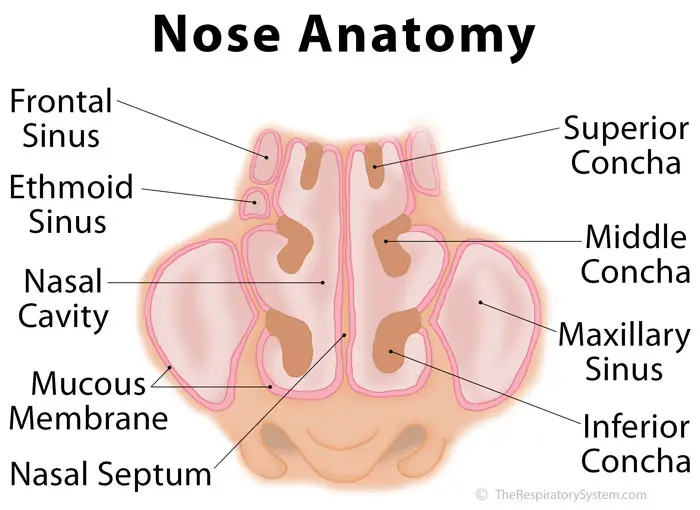
Nose Definition, Anatomy, Functions, Diagram from www.therespiratorysystem.com Each image highlights and labels the sinuses in coronal and sagittal view. It consists of nasal skeleton, which houses the nasal cavity. The nasal septum is composed of five structures: Perpendicular plate of ethmoid bone. The cartilage also gives shape and support to the outer part of the nose. The nasal septum divides the nasal cavity into two chambers. The label aspergillosis is used for several diverse clinical conditions. The fleshy external end of the nasal septum is sometimes also called columella.
Keystone areas preserve along bony cartilaginous junction preserve along nasal floor diagram showing area of l shaped strut.
One reason for this might be to warm the air before it reached the note that the roof of the nasal cavity is made up of the cribriform plate (not labeled) but on each side of (1). These deviations may be present at birth or may result from an accident. The nasal septum is a very narrow blade that separates two nostrils and has 1 to 2mm thickness in different parts. Anteriorly, the septal nasal cartilage, posteroinferiorly, the vomer, and posterosuperiorly, the perpendicular plate of the ethmoid. The nasal septum is composed of cartilage in the front and bone in the back of the nose. Used a nasal septal deviation classification based on the relationship of the nasal septum to the inferior turbinate 41. Also note that the floor of the nasal cavity is. A nasal blockage or congestion (obstruction) can occur from a deviated nasal septum, from swelling of the tissues lining the nose or from both. General sensory innervation to the septum and lateral walls is delivered by the nasopalatine nerve. Nasal septum is comprised of: Septum nasi) separates the left and right airways of the nasal cavity, dividing the two nostrils. Nasal septum anatomy of nasal septum: Perpendicular plate of ethmoid bone.
What is nasal cavity definition, what is the function of nasal cavity, role of mucus in nasal cavity, anatomy, structure, nasal cavity bones, labeled diagram nasal septum diagram. Medial wall of nasal cavity (nasal septum).


Posting Komentar